
Eye Surgery
Expert eye care services for various conditions and surgeries.


Cataract Surgery
Cataract surgery is a medical procedure to remove a cataract, a clouding of the lens in the eye that affects vision. Here's an overview:
Types of Cataract Surgery
1. Phacoemulsification: A modern technique using ultrasonic waves to break up the cataract.
2. Extracapsular cataract extraction (ECCE): A traditional method removing the lens in one piece.
3. Laser-assisted cataract surgery (LACS): Using a laser to create incisions and break up the cataract.
Procedure
1. Pre-operative evaluation: Assessing the cataract and overall eye health.
2. Surgery: Removing the cataract and implanting an intraocular lens (IOL).
3. Post-operative care: Monitoring healing and adjusting medications.
Benefits
1. Improved vision: Restoring clear vision and reducing glare.
2. Quick recovery: Most patients experience rapid recovery.
3. Low risk: Generally safe, with minimal complications.
Considerations
IOL selection: Selecting the right lens for optimal vision.
Monofocal IOLs
1. Fixed focus: One focal point for distance, intermediate, or near vision.
2. Commonly used: Suitable for most cataract patients.
Multifocal IOLs
1. Multiple focal points: Allows for clear vision at multiple distances.
2. Reduced dependence on glasses: Suitable for patients seeking reduced dependence on corrective eyewear.
Accommodating IOLs
1. Dynamic focusing: Moves with eye movements to provide clear vision at various distances.
2. More natural vision: Mimics the natural lens's ability to focus.
Toric IOLs
1. Astigmatism correction: Designed to correct astigmatism, providing clear distance vision.
2. Reduced astigmatism: Suitable for patients with significant astigmatism.
Extended Depth of Focus (EDOF) IOLs
1. Extended range of vision: Provides a range of clear vision, reducing dependence on glasses.
2. Intermediate and distance vision: Suitable for patients prioritizing intermediate and distance vision.


Glaucoma Treatment


Lasik Treatment
LASIK (Laser-Assisted In Situ Keratomileusis) is a popular and effective laser eye surgery that can correct vision problems like nearsightedness, farsightedness, and astigmatism—often giving you freedom from glasses or contact lenses.
⏱️ "15-Minute" LASIK – What Does That Mean?
Yes, LASIK is often referred to as a “15-minute” procedure. Here's why:
The entire LASIK procedure typically takes about 15 minutes per eye, sometimes even faster.
The actual laser correction part lasts just 20 to 60 seconds per eye, depending on the correction needed.
Most of the time is spent in preparation and ensuring proper alignment.
🧑⚕️How It Works:
Anesthetic eye drops numb your eye.
A femtosecond laser or microkeratome creates a thin flap in your cornea.
An excimer laser reshapes the cornea to correct vision.
The flap is repositioned—no stitches required.
✅ Benefits:
Quick and painless (some pressure, no sharp pain)
Fast recovery: Most people see improvement within a day.
Long-lasting results
Can correct most refractive errors
⚠️ Things to Know:
Not everyone is a candidate. You’ll need:
Stable vision for a year or more
Healthy corneas
No severe dry eyes or other eye conditions
Temporary side effects: glare, halos, dry eyes
Some people may still need reading glasses later in life (presbyopia)
👁️🗨️ Alternatives:
PRK (surface laser, for thinner corneas)
SMILE (minimally invasive)
ICL (Implantable Contact Lenses)
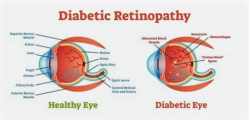
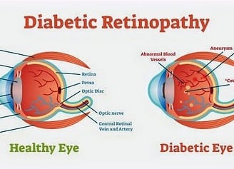
Diabetic Retinopathy is an eye condition that affects people with diabetes, and it's one of the leading causes of blindness in adults. It happens when high blood sugar levels damage the blood vessels in the retina, the light-sensitive tissue at the back of the eye.
There are two main stages:
Non-proliferative diabetic retinopathy (NPDR):
Early stage.
Blood vessels in the retina weaken and may leak fluid or blood.
You might not notice symptoms right away.
Can cause blurry vision or dark spots.
Proliferative diabetic retinopathy (PDR):
More advanced stage.
New, fragile blood vessels grow in the retina (proliferation) and can bleed into the eye.
Can lead to scarring, detached retina, and vision loss.
Common symptoms:
Blurred or fluctuating vision
Dark or empty areas in your vision
Floaters (small dark shapes or spots)
Vision loss (in more severe cases)
Risk factors:
Poorly controlled blood sugar
High blood pressure
High cholesterol
Duration of diabetes (the longer you’ve had it, the higher the risk)
Diagnosis:
Dilated eye exam: An ophthalmologist looks for signs of damage.
Fluorescein angiography or OCT (Optical Coherence Tomography) to get detailed images of the retina.
Treatment:
Laser therapy (photocoagulation) to seal leaking blood vessels
Anti-VEGF injections (like Avastin, Lucentis, or Eylea) to reduce blood vessel growth
Vitrectomy: Surgery to remove blood or scar tissue from the eye
Strict blood sugar and blood pressure control
Diabetic Retinopathy
Pediatric eye care
Pediatric eye care is a specialized branch of ophthalmology focused on the eye health and visual development of children, from infancy through adolescence. It includes the prevention, detection, and treatment of eye conditions that can affect children's vision.
Here’s an overview of what it typically involves:
👁️ Common Pediatric Eye Conditions:
Refractive Errors (need for glasses)
Myopia (nearsightedness)
Hyperopia (farsightedness)
Astigmatism
Strabismus (misaligned eyes)
One or both eyes may turn in, out, up, or down.
Amblyopia ("lazy eye")
One eye doesn’t develop proper vision. Often linked to strabismus or refractive errors.
Congenital Eye Disorders
Cataracts, glaucoma, ptosis (droopy eyelid), etc.
Blocked Tear Ducts
Common in infants and usually resolves on its own or with gentle massage.
Eye Injuries
More frequent in kids due to sports and play.
👩⚕️ Pediatric Eye Exams:
Eye exams for children are different from adult exams. They are adapted to be fun and interactive, using pictures, lights, and toys to keep a child’s attention.
Recommended timeline:
Newborn: Basic eye screening in the hospital
6–12 months: Comprehensive exam, especially if high-risk
3–5 years: Eye exam to detect conditions like amblyopia
Before starting school (around 5–6 years) and then every 1–2 years
🛠️ Treatments:
Eyeglasses or contact lenses
Patching therapy (for amblyopia)
Eye exercises or vision therapy
Surgical intervention (for strabismus, cataracts, etc.)
👶 Signs Parents Should Watch For:
Eye rubbing or squinting
Tilting the head to see better
Poor hand-eye coordination
Sitting too close to screens or books
One eye turning in or out
Frequent headaches or eye fatigue


Computer Vision Syndrome (CVS)
Computer Vision Syndrome (CVS)—also known as digital eye strain—refers to a group of eye and vision-related problems that result from prolonged use of digital screens like computers, tablets, e-readers, and smartphones.
🔍 Causes of CVS
Extended screen time
Poor lighting or glare
Improper viewing distance or angles
Uncorrected vision problems
Blinking less while staring at screens
👁️ Common Symptoms
Eye strain or discomfort
Dry eyes
Blurred or double vision
Headaches
Neck and shoulder pain
Difficulty focusing
🧠 How It Happens
When you're staring at a screen, your eyes have to work harder:
Constantly shifting focus
Moving back and forth across text
Adjusting to screen glare and contrast
Blinking rate drops significantly → leads to dry eyes
✅ Tips for Prevention and Relief
1. 20-20-20 Rule
Every 20 minutes, look at something 20 feet away for at least 20 seconds.
2. Blink More
Make a conscious effort to blink often to keep your eyes moist.
3. Screen Position
Keep the top of the screen at or slightly below eye level, 20–28 inches from your eyes.
4. Reduce Glare
Use anti-glare screens or filters, adjust lighting, and clean screens regularly.
5. Adjust Display Settings
Increase text size
Boost contrast
Reduce screen brightness
Use blue light filters or night mode
6. Use Proper Eyewear
Glasses with anti-reflective coating or blue light filtering can help.


Glaucoma treatment aims to lower intraocular pressure (IOP) to prevent or slow vision loss. Treatment options include:
Medications
1. Eye drops: Various types, such as prostaglandin analogs, beta-blockers, and alpha agonists.
2. Oral medications: In some cases, oral medications like acetazolamide may be prescribed.
Laser Therapy
1. Trabeculoplasty: Improves drainage in the eye.
2. Iridotomy: Creates a hole in the iris to improve drainage.
Surgery
1. Trabeculectomy: Creates a new drainage path.
2. Glaucoma drainage devices: Implants to facilitate drainage.
3. Minimally invasive glaucoma surgery (MIGS): Less invasive procedures with quicker recovery.
Lifestyle Changes
1. Regular exercise: May help lower IOP.
2. Healthy diet: A balanced diet rich in fruits, vegetables, and omega-3 fatty acids.
3. Monitoring: Regular eye exams to track IOP and adjust treatment.
Dr. Nanaware's expertise in eye surgery transformed my vision. Highly recommend his services for eye care!
Sanjay Mehta


★★★★★